|
Virtual Evaluation of the Accuracy of Fit and Trueness inMaxillary Poly(etheretherketone) Removable Partial DentureFrameworks Fabricated by Direct and Indirect CAD/CAMTechniques
|
|
|
Enas Elhamy Negm, BDS , Faten Ahmed Aboutaleb, PhD, & Ahmed M. Alam-Eldein, PhD
|
|
|
Department of Removable Prosthodontics, Faculty of Dentistry Tanta University, Tanta, Egypt
|
|
|
enas.negm@yahoo.com
|
|
|
68
|
|
|
Abstract
Purpose: To compare the accuracy of fit and trueness of maxillary poly(etheretherketone) (PEEK) removable partial denture (RPD) frameworks fabricated by direct and indirect computer-aided design/computer-aided manufacturing
(CAD/CAM) techniques.
Materials and Methods: A definitive maxillary class I master epoxy model was duplicated to create a stone cast. The cast was scanned, and an RPD was designed. Standard tessellation language (STL) data were used to fabricate 20 identical RPD frameworks using two CAD/CAM techniques: direct milling of PEEK and indirect additive manufacturing (resin printing combined with PEEK thermopressing using the lost-wax technique). All the frameworks for each technique (n = 10) and the reference cast were scanned. To assess the accuracy of fit, a color map was constructed using metrology software, and the misfit (distance between each framework and the reference cast) was measured at 25 standardized points. To assess the overall trueness, each framework STL file was superimposed over the original design’s STL file, and the average deviation was recorded in microns. Data were statistically analyzed using t-test.
Results: Color mapping showed distinct pressure areas in the anterior and posterior straps of the major connector in the indirect technique, while a more uniform distribution of the color map was observed in the direct technique. A significant difference was found between the two techniques regarding the overall accuracy of fit. Compared with the indirect technique, the milled frameworks showed significantly better overall trueness (p < 0.001).
Conclusions: Although a significant difference in the overall fit accuracy was noted between both techniques, the fit was an acceptable clinical fit. The fabrication method affects the fit in the anterior and posterior strap areas. The frameworks of the direct technique revealed better overall trueness values compared with the indirect technique.
|
|

|
Locator versus Equator Attachment Used with Mandibular Implant Retained Overdenture: An In-Vitro Study
|
|
|
Rana Osama Mohamed El-Bahrawy,Zeinab Ahmed El-shourbagy,Safa’a Al-Sayed Asal
|
|
|
prosthodontic department tanta university
|
|
|
ranabprosth@gmail.com
|
|
|
69
|
|
|
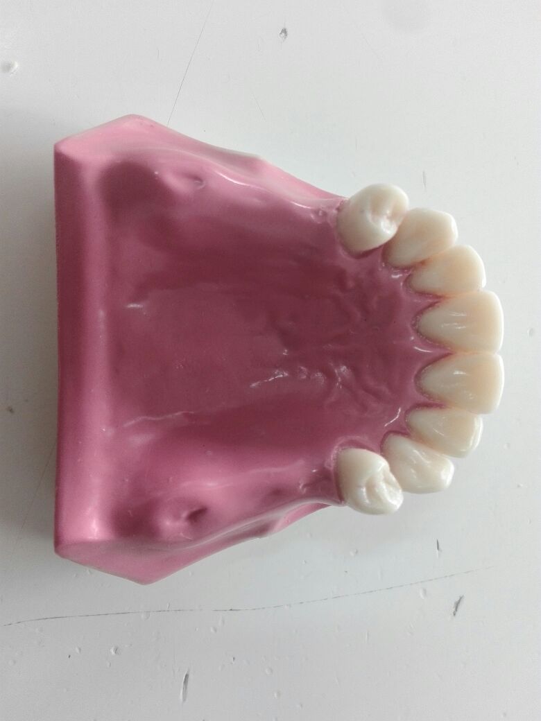
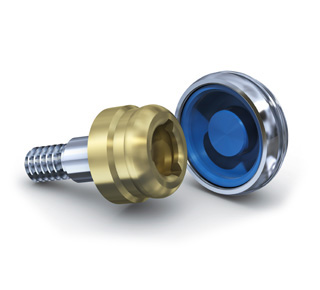
- Purpose: Compare retention and wear characteristics of Locator and OT equator attachments used in two implant retained mandibular overdenture. Materials and Methods: Fourteen readymade mandibular epoxy resin models were used as well as four opposing maxillary epoxy models. Two implant fixtures with 4 mm diameter and 11mm length were installed in the canine area. Group (I) received Locator abutments with white nylon inserts while Group (II) received OT equator attachments wit clear nylon inserts. The final constructed mandibular overdentures were subjected to 240,000 cycle of bi-axial chewing simulating cycles and 1080 manual insertion-removal cycles. The retention of the samples were measured using Universal Testing Machine at the beginning of the study and after 3,6,9,12 months of functional simulation. Wear features in the nylon inserts were also observed by USB digital microscope with built in camera at the beginning of the study and at 3,6,9,12 months. Results: The Locator attachments showed 31.89% loss of initial retention after one year of simulated function compared to 46.44% loss of OT equator retention. Notable wear and surface alterations were visible under photomicrograph on nylon inserts of both attachments. However, both systems nevertheless fulfilled the minimum retention requirements during simulation of one year functional life. Conclusion: Locator attachment system showed superior retentive properties when compared to OT equator. Statistically significant differences in retention develop between the two systems with OT equator undergoing a greater loss of retention than Locator
|
|

|
The Effect of Storage Times and Temperature Degrees of Cleansing Solutions on Retention of Overdenture Bar Attachments
|
|
|
Bahaa El-Deen Mohamed Abd el-Azeem,Ahmed Mohamed Alam-Eldein and Mohamed Maamoun El-Sheikh
|
|
|
tanta university
|
|
|
bahaa.mohamed@dent.tanta.edu.eg
|
|
|
74
|
|
|
Purpose: The aim of this in-vitro study was to examine the effect of storage times and temperature degrees of cleansing solutions on retention of overdenture bar attachments.
Materials and methods: 120 yellow hader bar clips were divided into two groups according to type of cleansing solution(Tape water and Corega), each group divided into 3 subgroups 20 each according to degree of temperature (A-20°C, B-37°C, C-60°C), each subgroup divided into 2 equal numbers according to storage time (1/2hour, 1hour). Each tested specimen consist of two parts, first part(block A) contained of two implants and the bar in rectangular acrylic resin block (2 × 1 × 1 inch)and second part (block B) contained of yellow hader clip and metal housing. Retention force was measured using universal testing machine and wear of the clips were checked before and after immersion in cleansing agents, the clips were observed under the microscope. The peak load-to-dislodgement of the specimens after each pull was documented, the data were collected and analyzed by T student-test and analysis of variance (ANOVA) and followed by tukey's HSDtest when P value ≤0.05 considered significant.
Results: Immersion in water and Corega at 60°C for ½ hour and 1 hour per day significantly increase the retention of bar attachment p≤0.05.While immersion at 20°C for ½ hour and 1 hour per day significantly decrease the retention of bar attachment p≤0.05 but at 37°C for ½ hour and 1 hour per day in water significantly decrease the retention of bar attachment p≤0.05 and at 37°C for ½ hour and 1 hour per day in Corega significantly increase the retention of bar attachment p≤0.05.SEM revealed surface changes in plastic clips after immersion in all groups.
Conclusions: Using denture cleansing solutions (Corega and water) at 60°C increase the retention value of bar attachments.
|
|
|
Stress Analysis of Short Implants with Different Diameters in Maxillary Bilateral Distal Extension Bases
|
|
|
Amr Azab Abd-Elfattah, Said M.Abd-Allah and Ali M. El-Sheikh
|
|
|
Tanta university
|
|
|
amr.azab@dent.tanta.edu.eg
|
|
|
78
|
|
|
Purpose: The aim of this study was to evaluate stress distribution around short implants with different diameters in maxillary bilateral distal extension bases. Materials and methods: Strain gauge technology was used for strain analysis for two different diameters of short implant. On the seven epoxy resin models, two different short implant diameters were used as follow: Group A; On one side of the model, two implants with 4.8 mm diameter and 5 mm length were used. One implant was placed in the second premolar region and the other was placed in the second molar region. For group B; Implants with 6.2 mm diameter and 5 mm length were used. For both groups, ceramo-metal screw retained fixed partial dentures were constructed on the transmucosal abutments. Stresses were measured at the buccal and lingual side of each implant under 200 N vertical load and 40 N oblique load. Data were collected, tabulated and statistically analyzed. Results: The present study revealed that minimum stresses were induced on vertical and oblique loads around the short implants that had wider diameters. Conclusions: within the limitation of this study, it could be concluded that, using short implants with wider diameter is a reliable method for restoring the distal extension bases.
|
|
|
Locator versus Equator Attachment Used with Mandibular Implant Retained Overdenture: An In-Vitro Study
|
|
|
Rana Osama Mohamed El-Bahrawy,Zeinab Ahmed El-shourbagy,Safa’a Al-Sayed Asal
|
|
|
prosthodontic department tanta university
|
|
|
ranabprosth@gmail.com
|
|
|
80
|
|
|
Purpose: Compare retention and wear characteristics of Locator and OT equator attachments used in two implant retained mandibular overdenture. Materials and Methods: Fourteen readymade: mandibular epoxy resin models were used as well as four opposing maxillary epoxy models. Two implant fixtures with 4 mm diameter and 11mm length were installed in the canine area. Group (I) received Locator abutments with white nylon inserts while Group (II) received OT equator attachments wit clear nylon inserts. The final constructed mandibular overdentures were subjected to 240,000 cycle of bi-axial chewing simulating cycles and 1080 manual insertion-removal cycles. The retention of the samples were measured using Universal Testing Machine at the beginning of the study and after 3,6,9,12 months of functional simulation. Wear features in the nylon inserts were also observed by USB digital microscope with built in camera at the beginning of the study and at 3,6,9,12 months. Results: The Locator attachments showed 31.89% loss of initial retention after one year of simulated function compared to 46.44% loss of OT equator retention. Notable wear and surface alterations were visible under photomicrograph on nylon inserts of both attachments. However, both systems nevertheless fulfilled the minimum retention requirements during simulation of one year functional life. Conclusion: Locator attachment system showed superior retentive properties when compared to OT equator. Statistically significant differences in retention develop between the two systems with OT equator undergoing a greater loss of retention than Locator
|
|
|
Biomedical Waste Disposal: knowledge, Attitude and Practice among Dental Practitioner in Tanta city, Egypt
|
|
|
Ahmed ibrahime El dosoky
|
|
|
El dosoky AI
|
|
|
ahmed_ibrahime@dent.tanta.edu.eg
|
|
|
221
|
|
|
Background: Dental waste is a risky division of biomedical waste (BM). Haphazard disposal of biomedical waste creates a significant threat to environment and human health. It has been observed that the guidelines for proper management of dental waste are not adopted and not up to the prescribed standard in most of the dental health facilities. So, proper management of biomedical waste turn out to be a significant concern for both the dental and general community. Effective management of biomedical waste is not only a legal necessity but also a social responsibility.
Aim: The aim of this study is to assess the knowledge, attitude and practice of biomedical waste generation, hazards, and legislation among a group of dental practitioner in Tanta city.
Methodology: A cross‑sectional study was conducted in 200 practicing dentists in Tanta city. A self‑structured questionnaire was used to obtain required data. The questionnaire was divided into three sections. The first section of the questionnaire contained questions regarding knowledge of BM waste generation, hazards, and legislation, whereas the second section contained questions regarding the level of awareness on BM waste management practice, and the third section contained questions regarding attitude/behavior toward BM waste.
Results: Of all study participants, 120 (60%) were males and 80 (40 %) were females. Among study participants, 89%knew about BM waste generation and legislation, whereas 5.5 % each did not know and were not aware of it. 80% of study sample would like to attend voluntarily programs that enhance and upgrade their knowledge about waste management.
Conclusion: There is a good level of knowledge and awareness about BM waste generation hazards, legislation, and management among health care personnel in Tanta city. Keywords: Attitude, practice, biomedical waste disposal, knowledge, dental practitioner
|
|
|
Awareness of Employees and Non-Medical Students at Tanta University toward Oral Cancer
|
|
|
Basant H. Abozaid, Nora A. Shalaby, Yomna S. Abd El-Aziz, Houssam Hebeish, Iman S. Elshamy, Madonna A. Morcos, Ahmed S. Rageh, Samar M. Amer, Mohamed A. Shaheen, Ranya W. Shehabeldeen, Esraa E. Elmalky, Asmaa M. Elsersy, Aya A. Dndn, Munir G. Abdel-Fattah
|
|
|
Faculty of Dentistry, Tanta University
|
|
|
basant_abozeed@dent.tanta.edu.eg
|
|
|
245
|
|
|
Worldwide, oral cancer burden is progressively rising, greatly influencing morbidity and mortality rates. The 5-year survival rates remain low typically due to delayed discovery until advanced stages. In addition, patient’s own perception of the disease in its early stages is usually subtle, since oral cancer frequently progresses without remarkable symptoms. Several studies highlighted the shortage of public awareness toward risk factors, signs and symptoms of oral cancer. There is a demanding need for assessing the level of oral cancer awareness among the public in order to create a positive attitude toward oral cancer early detection approaches. A cross-sectional questionnaire-based study was handed by third year dental students at Faculty of Dentistry, Tanta University to 110 randomly selected participants, 72 of them were employees at faculty of Dentistry, Tanta University and the remaining 38 were undergraduate non-medical students studying at faculties of Education, Engineering, Arts, Law, and Commerce, Tanta University. The present study was reviewed by the Tanta University, Faculty of Dentistry, Research Ethics Committee. Introductory section was added describing aim and benefit of the study in addition to voluntary participation in the research. All the participants didn’t receive any prior education about oral cancer. The majority underestimated the incidence and extent of oral cancer problem in Egypt. They passed over several oral cancer risk factors as well as clinical features including the most common site and different oral mucosal signs. The present study indicates the necessity for designing population-based awareness campaigns to raise the public knowledge about risk of oral cancer which would greatly impact the disease prognosis.
|
|
|
In Vitro Comparative Study of two Different Thermoplastic Resin Clasp Materials
|
|
|
Dena Mohey El-deen El-sayed Hamed, Nabeel M. El-Gendy, Usama Mahmoud Abdel Karim and Fadel El Said Abd El Fattah.
|
|
|
Facutly of Dentistry, Tanta university
|
|
|
dina_hamed@dent.tanta.edu.eg
|
|
|
258
|
|
|
Objectives: The objective of this study was to evaluate the retention, the flexural strength and modulus of two clasp materials (Acetal resin and Valplast Materials and Methods: For the retention test, on two standardized premolar metallic models, sixty-four Akers clasps were fabricated and classified into two groups, including thirty-two clasps for each material (Acetal and Valplast), Each group was divided into two subgroups A and B (sixteen clasps with 2mm thickness and sixteen with 3mm thickness). Each subgroup was subdivided into two divisions (eight clasps engaging 0.02 inch and eight clasps engaging 0.03 inch undercut). Each clasp was subjected to 1500, 3000, 4500, 6000 and 7500 cycles of insertion/removal test. Sixteenspecimens were fabricated for flexural strength and modulus tests (eight specimens for each material) with dimensions of (64×10×2.5)mm following ISO1567. These specimens were subjected to three-point bending test. Data were statistically analyzed using student T- test (at the 0.05 level of significance). Results: Acetal clasps of both dimensions had significantly higher retentive strength, flexural strength and modulus than Valplast clasps. Clasps engaging 0.02 inch undercut showed significantly less retention than clasps engaging 0.03 inch undercut also, Clasps with 2mm thickness showed significantly less retention than that of 3mm thickness. All clasps exhibited significant decrease in retentive strength from the first period of cyclic loading to the end of the cycling. Conclusion: There was significant difference in retentive strength, flexural strength and modulus between both materials. Keywords: Acetal, Valplast, retentive strength, cyclic loading, flexural strength and modulus.
|
|
|
Comparison of two different feeding methods for cleft palate babies
|
|
|
Amal Abed Elaziez Salem .lecturer in prosthodontic department Faculty of Dentistry, Tanta University
|
|
|
Tanta University
|
|
|
amal.salim@dent.tanta.edu.eg
|
|
|
260
|
|
|
Statement of problem
Clefts palate are among the most common congenital malformations world wide.A child born with cleft palate may experience difficulties while feeding. Nasal regurgitation and choking are common in infants with cleft palate because of inability of the palate to separate the nasal and oral cavities.
Purpose
The purpose of this study was to compare between flexible and rigid acrylic feeding appliance
Material and methods
Ten cleft palate neonates were selected from the outpatient Prosthetic clinic of the Faculty of Dentistry, Tanta University. Two different feeding appliances were constructed for each baby .No attempt was made to enforce one particular method of feeding; on the contrary, parents were encouraged to find the method which best suited their individual baby by a process of trial and error.
Results
Rigid acrylic feeding plate was significantly more effective statistically than flexible appliance.
Conclusions
Acrylic rigid feeding plate was more effective in overcoming feeding problems associated with cleft palate than flexible appliance
|
|

|
Relationship between Shear Bond Strength and Fluoride Content of Fluoridated Teeth as Stated by Scanning Electron Microscopy and X-Ray Fluorescence Analysis
|
|
|
Samy M El-Saftya*, Tasneem A Almohamadyb
|
|
|
a Lecturer, Department of Dental Biomaterials, Faculty of Dentistry, Tanta University, Tanta 31527, Egypt. b Orthodontist, El-Santa Central Hospital, El-Santa, Gharbia, Egypt.
|
|
|
drelsafti@yahoo.com
|
|
|
1399
|
|
|
Objectives: To study the effect of fluoride treatment on shear bond strength (SBS) of orthodontic brackets and to investigate the relationship between SBS and fluoride content of fluoridated teeth by means of scanning electron microscopy (SEM) and X-ray fluorescence (XRF) analysis. Materials and methods: A total of one hundred sound maxillary premolars were thoroughly cleaned and randomly assigned to 5 groups (n=20): control without fluoride treatment and four groups fluoridated with Profluorid® Varnish 5% NaF and stored in artificial saliva for varied periods: 1, 3, 5 and 7 days. After completing the predetermined storage time, specimens were subdivided into two subgroups (n=10); ten specimens were used for SBS testing. the other specimens were submitted to XRF to study fluoride content. Phosphoric acid 37% and a resin adhesive Transbond XT were used to bond the ceramic brackets to the teeth. Debonding was carried out in a universal testing machine. After debonding, a stereomicroscope determined the type of fracture of each specimen and scores of adhesive remnant index. SEM investigation was conducted on representative specimens of debonded tooth surfaces of groups in SEM. XRF analysis was carried out using AXIOS, WD-XRF Sequential Spectrometer. Crowns of specimens of each subgroup were cut, ground and pressed by the aid of binding wax to form disc-shaped specimens that were submitted to the XRF analysis. Significance level was established. Results: statistically significant differences were found between studied groups in both SBS values and ARI scores. SEM examination showed that fluoride layer acted as a barrier hindering proper etching of tooth surface that resulted in poorer bonding compared to control group. Longer storage, however, caused fluoride layer to be washed away giving better bonding. XRF analysis revealed greater fluoride content of groups with shorter storage time compared to those with longer storage.
|
|

|
Maxillary Molar Distalization by A Modified Palatally Anchored Expander
|
|
|
Mohamed Elsaharty , Neven Fakhry , Atia Abd Elwareth Yousif ,Ahmed Ashour and Safaa Ghobashy
|
|
|
Assistant Lecturer of Orthodontics, Faculty of Dentistry, Tanta University Lecturer of Orthodontics, Faculty of Dentistry, Tanta University Assistant Professor of Orthodontics, Faculty of Dentistry, Tanta University Resident Orthodontist,Faculty of Dentistry, Tanta University Professor of Orthodontics, Faculty of Dentistry, Tanta University
|
|
|
mohamed.elshaikh@dent.tanta.edu.eg
|
|
|
1401
|
|
|
Objectives:The presentstudy was conducted to achieve bodily molar distalization by a modified skeletally anchored palatal expander. Materials and methods: Four patients (3 boys and a girl; mean age 11.3 years) were treated by a modified skeletally anchored palatal expander. All patients were dentally Class II molar relation with deep overbite and normal or sagittally directed growth pattern. Lateral cephalograms were taken and analyzed before and after molar distalization. Maxillary molar distalization was carried out bya modified HYREX palatal expander anchored to 2 anteriorly positioned palatal miniscrews (2.1 mm in diameter and 11 mm in length). The appliance was activated twice weeklyto exerta force of 90 gm/activation. Results: The appliance was able to move the maxillary first molars distally by an average of 5.23+ 1.23 mm without tipping and Class I molar relation was obtained within a period of 4.63 + 1.2 months. Slight extrusion of the maxillary molars was observed which was reflected on the opening rotation of the mandible, as the FH/MP angle was increased by 1.250 + 0.230 and the increase in the Y axis by 1.930 + 0.230 . A marked improvement of the deepbite was observed. Conclusion: The modified palatally anchored expander was an effective treatment alternative for the management of Class II non extraction cases by maxillary molar distalization.
|
|
|
THE EFFECT OF RECYCLING AND REPEATED RECYCLING WITH Er:YAG LASER ON SHEAR BOND STRENGTH AND SURFACE CHARACTERISTICS OF STAINLESS STEEL ORTHODONTIC BRACKETS
|
|
|
Safa B. M. Alawy, Eman M. El Shourbagy and Safaa A. Ghobashy
|
|
|
Faculty of dentistry, Tanta university
|
|
|
safa_basyouny@dent.tanta.edu.eg
|
|
|
1410
|
|
|
Aim of the work: The aim of this in vitro study was to evaluate the effects of recycling and repeated recycling with erbium: Yttrium aluminum garnet (Er:YAG) laser on shear bond strength and surface characteristics of stainless steel orthodontic brackets.
Materials and Methods: Ninety stainless steel brackets were bonded to ninety extracted premolars and were randomly divided into three groups of 30 teeth/each: group I (control), group II (brackets debonded and recycled one time using Er:YAG laser), and group III (brackets debonded and recycled two times using Er:YAG laser). Twenty samples from group II and III were rebonded again to the same re-prepared enamel. Sixty bonded samples (20/each group) were subjected to shear bond strength testing using universal testing machine. Ten debonded brackets from each group were examined under a scanning electron microscope to identify the surface characteristics of the brackets’ mesh.
Results: The results of this study demonstrated that the mean shear bond strength of all groups were more than that recommended for successful orthodontic bonding, there was no significant difference between group I and group II (P>0.05). Repeated recycled brackets (group III) had significantly lower shear bond strength (P≤0.001) than other groups. Scanning electron microscopic analysis showed maximal removal of adhesive from the brackets recycled one time with Er:YAG laser with well-defined lattice structure, and no damage to the bracket base. There was incomplete removal of the adhesive from repeated recycled brackets, flattening and distortion were observed in two samples.
Conclusion: Er:YAG laser recycled brackets can be used as an alternative to new brackets in case of bond failure which might provide cost reduction. However, in case of second bond failure to the same bracket, it may be preferred to use a new one instead of reusing it for second time.
|
|

|
REMINERALIZATION EFFICIENCY OF CHICKEN EGG SHELL POWDER (CESP) ON ARTIFICIALLY INDUCED DENTAL EROSION IN PRIMARY TEETH: AN IN-VITRO STUDY
|
|
|
Eman Mohamed Sobhy El-Bahrawya,* ,Ahmed Ibrahime El- Dosokyb
|
|
|
aDental Biomaterials Dept., bOral Health & Preventive Dentistry, Faculty Of Dentistry, Tanta University, Tanta, Egypt
|
|
|
dreman1978@yahoo.com
|
|
|
1424
|
|
|
Several cases of tooth erosion are attributed to oral administration of liquid oral medications. Chicken egg shell powder (CESP) has gained much concern in modern dentistry for having anti-erosive properties. The aim of this study was to evaluate the effect of CESP on the surface roughness and microhardness of primary teeth enamel eroded by antihistaminicsyrup. Twenty exfoliated/ extracted primary teeth were collected for the study. Then, each tooth was sectioned buccolingually into two halves, as study group and as control. Specimens of both groups were immersed in antihistaminic syrup for 30min twice daily for 12 days to produce erosive enamel lesions. Study group specimens were treated with the CESP solutiononce daily 3min for 10 consecutive days while control group specimens remained untreated. The specimens were stored in artificial saliva between the erosive and remineralizing cycles, then were evaluated for surface roughness using surface profile gauge and surface microhardness using Vickers microhardness tester. The mean value of surface roughness was significantly increased after erosion with antihistaminic syrup. The mean surface roughness of remineralized samples was significantly lower than that of the untreated samples. The mean value of surface microhardness was significantly decreased after erosion with antihistamine syrup. The mean surface microhardness of remineralized samples was significantly higher than that of the untreated samples. Treatment of eroded primary tooth enamel with CESP solution decreased surface roughness and improved surface microhardness in-vitro. Roughness and hardness testing are accurate and reliable methods to evaluate the enamel surface for demineralization and remineralization studies in vitro.
|
|

|
In Vivo Effect of Bioactive Glass with Fluoride on Enamel Surface Prior to Orthodontic Brackets Bonding
|
|
|
Hazem Magdy
|
|
|
Faculty of Dentistry, Tanya University
|
|
|
mohamed.elshaikh@dent.tanta.edu.eg
|
|
|
1425
|
|
|
To investigate the in vivo effects of NUPRO® extra care prophy paste on preventing enamel demineralization surrounding orthodontic brackets before bonding using Atomic Force Microscopy. This study was carried out on twenty patients scheduled to have maxillary premolars extracted as a part of orthodontic treatment. Participants in this study were divided into 2 equal groups according to the type of prophylaxis paste used before bonding utilizing split mouth technique. Group I (experimental side) n = 20 premolar in which NUPRO extra care phrophypaste was used in right side and Group II(controlside) n = 20 premolar in which conventional fluoride-free polishing paste was used in left side . The participants werefurther divided into anothertwo groups 10 per each according to the duration of the experiment into Group A and Group B for either 30 days or 60 days. Stainless steel brackets were bonded to upper premolars in all casesafter prophylaxis and T-loops stainless steel wires are engaged on brackets to increase plaque accumulation. After the brackets were debonded teeth were extracted and prepared for atomic force microscopic scanning. The mean average roughnessand total surface area were calculated for each sample. Comparison between groups was performed using one way ANOVA test with P ≤ 0.05. Group IA and Group IB yielded the least mean average roughness and total surface area than control groups (Group IIA and Group IIB). In conclusion, Single Application of NUPRO extra care phrophypaste prior to orthodontic bracket bonding provides reduction in enamel demineralization around orthodontic attachmentsforonly one month.
|
|

|
Efficiency of Intraoperative Fluorescence Imaging in Treatment of Patients with Bisphosphonate Related Osteonecrosis of the Jaw
|
|
|
Eslam Elhaliem Gharieb , Abdelfattah A. Sadakah , Mohammad A. Elshall , Elsayed M. Deraz
|
|
|
Faculty of Dentistry, Tanta University
|
|
|
eslam_gharieb@dent.tanta.edu.eg
|
|
|
1426
|
|
|
Purpose: to evaluate the accuracy of intraoperative fluorescence imaging indifferentiating vital from necrotic bone via incorporating doxycycline into mineralizingbone by the aid of (Oral ID™) device in patients with bisphosphonate relatedosteonecrosis of the jaws (BRONJ) disease. At Oral and Maxillofacial surgery department, Faculty of Dentistry, Tanta University. Prospective non controlled clinical study was carried outon twelve patients above 30 years old who had (BRONJ) disease. Intraoperative, after surgical exposure, visual fluorescence retention (VFR) and visualfluorescence loss (VFL) were documented on digital photographs. VFL appears dark(necrotic bone) showing a distinct contrast to surrounding normal pale greenfluorescence (vital tissue) , The necrotic bone was curetted under normal light using bone resected (Biopsy I), with usage of the device, more surgical resection of the affected VFL darkbone were done till VFL ceases the resected bone biopsy (Biopsy II), peripheral ostectomy of 2 mm of the compact bone (VFR) [taken as safety margin] (BiopsyIII).Post-operative, clinical evaluation of pain and wound healing. Histopathologicalevaluation was performed to detect necrotic and vital bone in all biopsies as well aspresence or absence of inflammatory cell infiltrate. Radiographic evaluation by CBCT. Results: The results show that all patients had complete relief of pain, complete healing of the soft tissuewas observed in ten patients, and two patients had partial soft tissue healing. Concerning histopathological evaluation, confirmed that image fluorescence is an effectiveIn complete eradication of necrotic bone from BRONJ lesions. In conclusion, the clinical and histopathological data suggested that the intraoperativefluorescence imaging is accurate in surgical management of BRONJ disease.
|
|

|
Clinical and Radiographic Evaluation of 3D Titanium Miniplate in Fixation of Anterior Mandibular Fractures
|
|
|
Ahmed Abd El LatifMosleh*; Mohamed M .Shoushan**; Mohamed M. SaadKhedr* and Ahmed S. Naguib*
|
|
|
* Oral and Maxillofacial Surgery Dept., ** Head of Oral and Maxillofacial Surgery Dept.,
|
|
|
ahmed_mosleh@dent.tanta.edu.eg
|
|
|
1427
|
|
|
purpose: the aim of this study was to evaluate the use of curved 2.0 mm 3-Dimensional strut titanium plate for fixation of anterior mandibular fractures (symphyseal or parasymphyseal). patients and methods: a prospective randomized clinical study was carried out in 12 adult patients with fractured symphyseal or parasymphyseal region of the mandible with well-defined inclusion and exclusion criteria. All patients were followedup for 6 months both clinically (for detection of neurosensory deficits of the lower lip, wound dehiscence, infection, segmental mobil¬ity, postoperative occlusion and degree of patient’s satisfactions with the performed treatment) and radiographically (for radiological evaluation of reduction and fixation). Results: twelve adult patients (9males and 3 females) were included in this work with one case (case no. 4) suffered from minor Occlusal discrepancy corrected by guiding elastics for ten days. Stability of fractured segments was achieved in all cases and examined clinically at all follow up periods (immediate postoperative, three months and six months) bimanually. One case suffered from wound dehiscence (case no.2) treated by daily irrigation with warm normal saline 0.9%, antiseptic mouth rinses. Immediate postoperative radiograph (panoramic x-ray-Axial and 3 dimen¬sional CT films) revealed no changes in position of reduced segments in horizontal and vertical direction of all cases. Radiographic examination revealed that the fracture line is difficult to be detected three months postoperatively and disappeared after six months.By the end of follow up period none of patients showed any signs of nonunion. conclusions: The 3-dimensional curved strut miniplate is considered as a better and easy sys¬tem to be applied for fixation of anterior mandibular fractures. But, there are some limitations to use in cases of oblique fractures and those fractures that involve the mental foramen, as well as there is excessive implant material because of the extra vertical bars
|
|

|
Effect of Different Root Canal Irrigation solutions on Bond Strength of Bioceramic-based Sealer
|
|
|
Abd-Elmoneim A. Elkalashy, Hatem A. Alhadainy, Walaa M. Ghoneim
|
|
|
Endodontics Department. Faculty of Dentistry, Tanya University
|
|
|
Abdelmoneim_elkalashy@yahoo.com
|
|
|
1429
|
|
|
Purpose: is to evaluate the possible effect of N-acetylcysteine (NAC), Sodium hypochlorite (NaOCl) and Chlorhexidine gluconate (CHX) on push-out bond strength of Endosequence BC sealer to radicular dentin.
Materials and methods: Forty extracted human single rooted premolars with one straight root canal were used, crowns of all teeth were removed to a root length of 15±1 mm, then roots were randomly divided into four equal groups (n=10) according to root canal irrigating solution, Group I; NAC solution, Group II; NaOCl solution, Group III; CHX solution and Group IV; saline solution as a control group. After working length determination, all canals were prepared using ProTaper rotary files up to #F3 using 5 ml of assigned irrigating solution after the use of each file. All root canals were obturated with gutta-percha cones in combination with Endosequence BC sealer. Three 2 mm thickness sections were obtained from each root representing coronal, middle and apical thirds and subjected to push-out test. Statistical analysis was performed using One Way Analysis of variance (ANOVA) at P-value ≤ 0.05.
Results: CHX recorded the highest mean value of push out bond strength at coronal and apical levels followed by NAC, NaOCl and saline solution. Coronal level displayed the highest mean bond strength value whereas the lowest one was recorded apically.
Conclusion: Irrigation with CHX and NAC solutions increased significantly bonding of Endosequence BC sealer to root canal dentin.
Keywords: Chlorhexidine gluconate, Endosequence BC sealer, N-Acetylcystine, Push-out bond strength, Root canal irrigation.
|
|
|